MRI Adrenal Gland
Indications for MRI Adrenal Glands
- Investigating the causes of hormonal imbalances
- Adrenal adenomas
- Adrenal tumours
- Cancers
Adrenal glands anatomy
The adrenal glands are small, triangular-shaped endocrine glands located on top of each kidney. Consisting of two distinct regions, the outer cortex and inner medulla, they play a crucial role in hormone production and regulation. The adrenal cortex synthesizes and secretes steroid hormones such as cortisol, aldosterone, and sex hormones. These hormones are vital for metabolism, blood pressure regulation, and reproduction. On the other hand, the adrenal medulla produces and releases catecholamines, including adrenaline and noradrenaline, which are involved in the body’s fight-or-flight response. The adrenal glands are highly vascularized and receive a rich blood supply, allowing for efficient hormone production and distribution throughout the body.

Contraindications
- Any electrically, magnetically or mechanically activated implant (e.g. cardiac pacemaker, insulin pump biostimulator, neurostimulator, cochlear implant, and hearing aids)
- Intracranial aneurysm clips (unless made of titanium)
- Pregnancy (risk vs benefit ratio to be assessed)
- Ferromagnetic surgical clips or staples
- Metallic foreign body in the eye
- Metal shrapnel or bullet
Patient preparation for MRI Adrenal Gland
- A satisfactory written consent form must be taken from the patient before entering the scanner room
- Ask the patient to remove all metal objects including keys, coins, wallet, cards with magnetic strips, jewellery, hearing aid and hairpins
- Ask the patient to undress and change into a hospital gown
-
Instruct the patient to hold their breath for the breath hold scans and breathe gently for the gated scans (it is advisable to coach the patient two to three times before starting the scan) - Claustrophobic patients may be accompanied into the scanner room e.g. by staff member or relative with proper safety screening
- Offer earplugs or headphones, possibly with music for extra comfort
- Explain the procedure to the patient
- Instruct the patient to keep still
- Note down the weight of the patient
Positioning for MRI Adrenal Gland
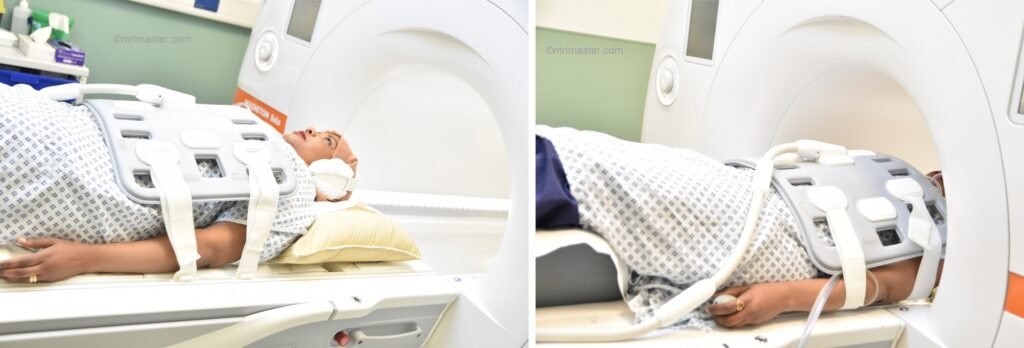
- Position the patient in supine position with head pointing towards the magnet (head first supine)
- Position the patient over the spine coil and place the body coil over the abdomen from the xiphoid process down to anterior superior iliac spine
- Securely tighten the body coil using straps to prevent respiratory artefacts
- Give a pillow under the head and cushions under the legs for extra comfort
- Centre the laser beam localiser over the level of lower intercostal border (i.e. L3 level)
Recommended MRI Adrenal Gland Protocols and Planning
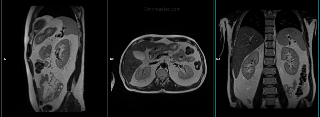
localiser
A three-plane T2 HASTE localizer must be taken initially to localize and plan the sequences. These fast, single-shot localizers usually have an acquisition time of less than 25 seconds, which is excellent for localizing abdominal structures.
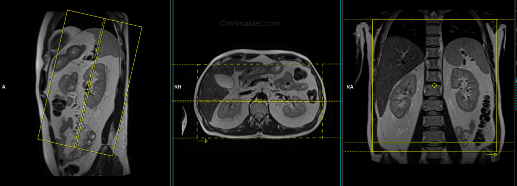
T2 tse breath hold (HASTE) coronal 3mm
Plan the coronal slices on the axial plane; angle the positioning block parallel to the midline along the right and left kidneys. Check the positioning block in the other two planes. An appropriate angle must be given in the sagittal plane, parallel to the long axis of the kidney. Slices must be sufficient to cover both kidneys anterior to posterior. Phase oversampling and, in the case of 3D blocks, slice oversampling, must be used to avoid wraparound artifacts. Instruct the patient to hold their breath during image acquisition. In our department, we instruct the patients to breathe in and out twice before the “breathe in and hold” instruction.
Parameters
| TR 5000-6000 | TE 150 | FLIP 150 | NXA 1 | SLICE 3MM | MATRIX 256×256 | FOV 300 | PHASE R>L | OVERSAMPLE 50% | IPAT ON |
T1 vibe DIXON breath hold coronal 3mm
Plan the coronal slices on the axial plane; angle the positioning block parallel to the midline along the right and left kidneys. Check the positioning block in the other two planes. An appropriate angle must be given in the sagittal plane, parallel to the long axis of the kidney. Slices must be sufficient to cover both kidneys anterior to posterior. Phase oversampling and, in the case of 3D blocks, slice oversampling, must be used to avoid wraparound artifacts. Instruct the patient to hold their breath during image acquisition. In our department, we instruct the patients to breathe in and out twice before the “breathe in and hold” instruction.
Parameters
TR 6-7 | TE 2.39 4.77 | FLIP 10 | NXA 1 | SLICE 4 MM | MATRIX 320×320 | FOV 320-350 | PHASE A>P | OVERSAMPLE 20% | BH YES |
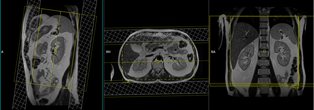
T1 VIBE DIXON 3mm axial BH pre GD(In-opposed phase and water sat)
Plan the axial slices on the coronal plane; angle the positioning block perpendicular to the thoracic vertebra. Check the positioning block in the other two planes. An appropriate angle must be given in the sagittal plane, perpendicular to the long axis of the kidney. Slices must be sufficient to cover both adrenals from the diaphragm down to the lower pole of the kidneys. Phase oversampling and, in the case of 3D blocks, slice oversampling must be used to avoid wrap-around artifacts. Using a saturation band on the top and bottom of the block will help to reduce artifacts from vascular pulsation and breathing. Instruct the patient to hold their breath during image acquisition.
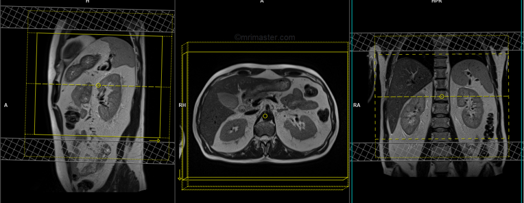
When planning the axial breath-hold scans, it is crucial to utilize the breath-hold vibe coronal sequence. This is because during inhalation, the diaphragm exerts downward pressure on the liver, causing a shift in its position from the initial localizer scans. Therefore, to accurately capture the desired imaging area of the liver, it is important to account for this positional change by utilizing the breath-hold vibe coronal sequence.

Parameters
TR 6-7 | TE 2.39 4.77 | FLIP 10 | NXA 1 | SLICE 3 MM | MATRIX 320×320 | FOV 320-350 | PHASE A>P | OVERSAMPLE 20% | BH YES |
T2 tse\HASTE breath hold 3mm axial
Plan the axial slices on the coronal plane; angle the positioning block perpendicular to the thoracic vertebra. Check the positioning block in the other two planes. An appropriate angle must be given in the sagittal plane, perpendicular to the long axis of the kidney. Slices must be sufficient to cover both adrenals from the diaphragm down to the lower pole of the kidneys. Phase oversampling must be used to avoid wrap-around artifacts. Using a saturation band on the top and bottom of the block will help reduce artifacts from vascular pulsation and breathing. Instruct the patient to hold their breath during image acquisition.
Note: T2 breath-hold sequences may have artifacts from the anterior abdominal fat motion. To reduce this artifact, it is highly recommended to use a right-to-left phase direction in these scans. However, this might not be possible in older generation scanners. In such cases, it is better to use a HASTE sequence in those scanners.

Parameters
| TR 5000-6000 | TE 150 | FLIP 150 | NXA 1 | SLICE 3MM | MATRIX 256×256 | FOV 280-300 | PHASE R>L | OVERSAMPLE 50% | IPAT ON |
T2 tse\HASTE fat sat breath hold axial 3mm
Plan the axial slices on the coronal plane; angle the positioning block perpendicular to the thoracic vertebra. Check the positioning block in the other two planes. An appropriate angle must be given in the sagittal plane, perpendicular to the long axis of the kidney. Slices must be sufficient to cover both adrenals from the diaphragm down to the lower pole of the kidneys. Phase oversampling must be used to avoid wrap-around artifacts. Using a saturation band on the top and bottom of the block will help reduce artifacts from vascular pulsation and breathing. Instruct the patient to hold their breath during image acquisition.
Parameters
| TR 6000-7000 | TE 150 | fatsat ON | NXA 1 | SLICE 3MM | MATRIX 256×256 | FOV 280-300 | PHASE R>L | OVERSAMPLE 50% | IPAT ON |
DWI epi 3 scan trace axial 3mm free breathing
Plan the axial slices on the coronal free-breathing localizer; angle the positioning block perpendicular to the thoracic vertebra. Check the positioning block in the other two planes. An appropriate angle must be given in the sagittal plane, perpendicular to the long axis of the kidney. Slices must be sufficient to cover both adrenals from the diaphragm down to the lower pole of the kidneys. Phase oversampling must be used to avoid wrap-around artifacts. Using a saturation band on the top and bottom of the block will help reduce artifacts from vascular pulsation and breathing. Instruct the patient to take shallow breathing during the acquisition.
Parameters
TR 6000-7000 | TE 90 | IPAT ON | NEX 3 5 8 | SLICE 3 MM | MATRIX 192X192 | FOV 200-250 | PHASE R>L | GAP 10% | B VALUE 0 |
T1 vibe DIXON breath hold Axial 3mm post contrast
Plan the axial slices on the coronal plane; angle the positioning block perpendicular to the thoracic vertebra. Check the positioning block in the other two planes. An appropriate angle must be given in the sagittal plane, perpendicular to the long axis of the kidney. Slices must be sufficient to cover both adrenals from the diaphragm down to the lower pole of the kidneys. Phase oversampling and, in the case of 3D blocks, slice oversampling must be used to avoid wrap-around artifacts. Using a saturation band on the top and bottom of the block will help to reduce artifacts from vascular pulsation and breathing. Instruct the patient to hold their breath during image acquisition.
Parameters VIBE dixon
TR 6-7 | TE 2.39 4.77 | FLIP 10 | NXA 1 | SLICE 3 MM | MATRIX 288×288 | FOV 320-350 | PHASE A>P | OVERSAMPLE 20% | BH YES |
T1 vibe DIXON breath hold coronal 3mm post contrast
Plan the coronal slices on the axial plane; angle the positioning block parallel to the midline along the right and left kidneys. Check the positioning block in the other two planes. An appropriate angle must be given in the sagittal plane, parallel to the long axis of the kidney. Slices must be sufficient to cover both kidneys anterior to posterior. Phase oversampling and, in the case of 3D blocks, slice oversampling, must be used to avoid wraparound artifacts. Instruct the patient to hold their breath during image acquisition. In our department, we instruct the patients to breathe in and out twice before the “breathe in and hold” instruction.
Parameters VIBE dixon
TR 6-7 | TE 2.39 4.77 | FLIP 10 | NXA 1 | SLICE 3 MM | MATRIX 288×288 | FOV 320-350 | PHASE R>L | OVERSAMPLE 40% | BH YES |

