MRI Brain Epilepsy (Seizure) Protocol
Indications for Epilepsy MRI scan
- Onset of generalised or unclassified seizures in the first year of life, or in adulthood
- Difficulty obtaining seizure control with first-line antiepileptic drugs (AEDs)
- Loss of seizure control, or a change in the pattern of seizures
- Onset of partial seizures, at any age
Contraindications
- Any electrically, magnetically or mechanically activated implant (e.g. cardiac pacemaker, insulin pump biostimulator, neurostimulator, cochlear implant, and hearing aids)
- Intracranial aneurysm clips (unless made of titanium)
- Pregnancy (risk vs benefit ratio to be assessed)
- Ferromagnetic surgical clips or staples
- Metallic foreign body in the eye
- Metal shrapnel or bullet
Patient preparation for Epilepsy MRI scan
- A satisfactory written consent form must be taken from the patient before entering the scanner room
- Ask the patient to remove all metal objects including keys, coins, wallet, cards with magnetic strips, jewellery, hearing aid and hairpins
- If possible provide a chaperone for claustrophobic patients (e.g. relative or staff )
- Offer earplugs or headphones, possibly with music for extra comfort
- Explain the procedure to the patient
- Instruct the patient to keep still
- Note the hight and weight of the patient
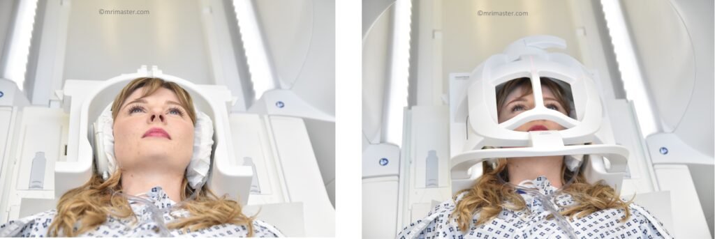
Positioning for Epilepsy MRI scan
- Head first supine
- Position the head in the head coil and immobilise with cushions
- Give cushions under the legs for extra comfort
- Centre the laser beam localizer over the glabella
Recommended Epilepsy MRI Protocols, Parameters, and Planning
Epilepsy MRI scan Localiser
A three-plane localizer must be taken at the beginning to localize and plan the sequences. Localizers are usually less than 25 seconds and are T1-weighted low-resolution scans.
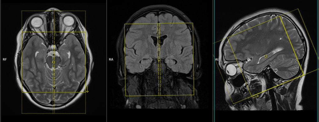
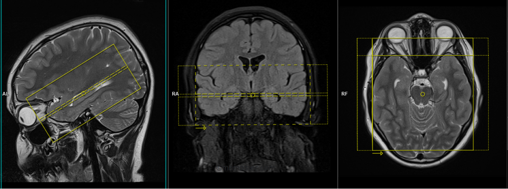
T2 tse axial
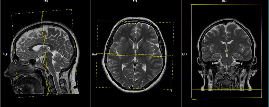
Plan the axial slices on the sagittal plane and position the block parallel to the genu and splenium of the corpus callosum. Verify the planning block in the other two planes and ensure that an appropriate angle is maintained in the coronal plane, making it perpendicular to the line of the midline of the brain and the 4th ventricle. Ensure that the number of slices is sufficient to cover the entire brain from the vertex to the line of the foramen magnum.
Parameters
TR 4000-5500 | TE 100-120 | SLICE 3MM | FLIP 130-150 | PHASE R>L | MATRIX 320X320 | FOV 210-230 | GAP 10% | NEX(AVRAGE) 2 |
T2 FLAIR axial
Plan the axial slices on the sagittal plane and position the block parallel to the genu and splenium of the corpus callosum. Verify the planning block in the other two planes and ensure that an appropriate angle is maintained in the coronal plane, making it perpendicular to the line of the midline of the brain and the 4th ventricle. Ensure that the number of slices is sufficient to cover the entire brain from the vertex to the line of the foramen magnum.
Parameters
TR 7000-9000 | TE 110 | FLIP 130 | NEX 2 | SLICE 3MM | MATRIX 320X320 | FOV 210-230 | PHASE R>L | GAP 10% | TI 2500 |
RESOLVE DWI axial
Plan the axial slices on the sagittal plane and position the block parallel to the genu and splenium of the corpus callosum. Verify the planning block in the other two planes and ensure that an appropriate angle is maintained in the coronal plane, making it perpendicular to the line of the midline of the brain and the 4th ventricle. Ensure that the number of slices is sufficient to cover the entire brain from the vertex to the line of the foramen magnum.
RESOLVE DWI: RESOLVE is an advanced technique used to obtain high-quality and high-resolution DWI (diffusion-weighted imaging) images, particularly in body regions affected by susceptibility artifacts. It offers sharp imaging with minimal distortions and provides excellent spatial resolution. RESOLVE is especially valuable for evaluating smaller lesions in a wide range of DWI and DTI (diffusion tensor imaging) examinations. By combining RESOLVE with Simultaneous Multi-Slice (SMS) acceleration, acquisition time can be significantly reduced, improving both speed and imaging capabilities. RESOLVE finds application in various clinical fields, including neurology, oncology, and pediatric imaging. Its outstanding balance between imaging speed and quality makes it superior to other diffusion-weighted imaging sequences. Benefits of RESOLVE include high-quality DWI and DTI, reduced susceptibility and blurring artifacts, insensitivity to motion-induced phase errors, lower specific absorption rate (SAR) compared to TSE-based methods, and compatibility with iPAT for faster scans and further reduced distortions.
Parameters
TR 7000-9000 | TE 70 | FLIP 130 | NXA 1 2 | SLICE 3MM | MATRIX 192X192 | FOV 210-230 | PHASE R>L | GAP 10% | B VALUE 0 |
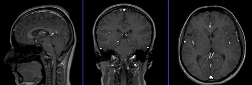
T1 SE coronal
Plan the coronal slices on the sagittal plane and angle the positioning block perpendicular to the line along the genu and splenium of the corpus callosum. Verify the planning block in the other two planes. Ensure that an appropriate angle is maintained in the axial plane, perpendicular to the midline of the brain. The number of slices should be sufficient to cover the entire brain from the frontal sinus to the line of the occipital protuberance.
Parameters
TR 500-700 | TE 15-25 | SLICE 3MM | FLIP 90 | PHASE R>L | MATRIX 304X304 | FOV 210-230 | GAP 10% | NEX(AVRAGE) 2 |
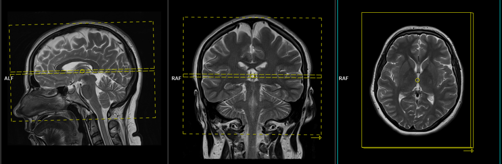
T2 tse sagittal
Plan the sagittal slices on the axial plane and position the block parallel to the midline of the brain. Verify the planning block in the other two planes. Angle the positioning block appropriately in the coronal plane, ensuring it is parallel to the line along the midline of the brain and the 4th ventricle. Make sure that the number of slices is sufficient to cover the entire brain from one temporal lobe to the other.
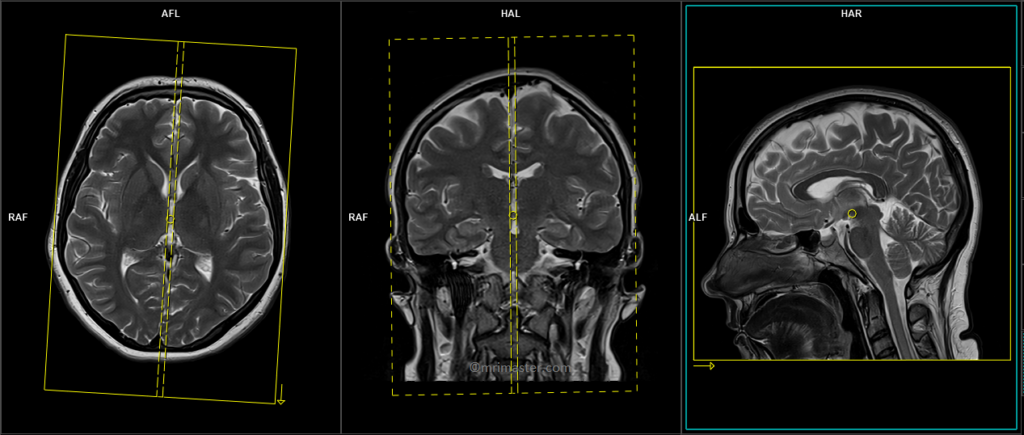
Parameters
TR 4500-6000 | TE 100-120 | SLICE 3MM | FLIP 130-150 | PHASE A>P | MATRIX 320X304 | FOV 210-230 | GAP 10% | NEX(AVRAGE) 2 |
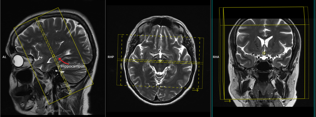
T2 tse coronal oblique 2mm ( epilepsy protocol)
Plan the coronal high-resolution slices on the sagittal plane and angle the planning block perpendicular to the long axis of the hippocampus. Check the planning block in the other two planes. An appropriate angle must be given in the axial plane, which is perpendicular to the midline of the brain. Ensure that the slices are sufficient to cover the entire temporal lobe

Parameters
TR 3000-4000 | TE 100-120 | SLICE 2 MM | FLIP 130-150 | PHASE A>P | MATRIX 320X320 | FOV 170-190 | GAP 10% | NEX(AVRAGE) 4 |
T1 tse coronal oblique 2mm ( epilepsy protocol)
Plan the coronal high-resolution slices on the sagittal plane and angle the planning block perpendicular to the long axis of the hippocampus. Check the planning block in the other two planes. An appropriate angle must be given in the axial plane, which is perpendicular to the midline of the brain. Ensure that the slices are sufficient to cover the entire temporal lobe
Parameters
TR 400-600 | TE 15-25 | SLICE 2 MM | FLIP 140 | PHASE R>L | MATRIX 320X320 | FOV 170-190 | GAP 10% | NEX(AVRAGE) 4 |
In children, it is strongly recommended to undertake 3-dimensional (3D) T1-weighted SPACE or SPGR images with 1mm slices in the coronal oblique plane (perpendicular to the hippocampus), including the entire brain from nasion to inion.
T1 SPACE coronal oblique 1mm
Plan the coronal high-resolution slices on the sagittal plane and angle the planning block perpendicular to the long axis of the hippocampus. Check the planning block in the other two planes. An appropriate angle must be given in the axial plane, which is perpendicular to the midline of the brain. Ensure that the slices are sufficient to cover the entire brain
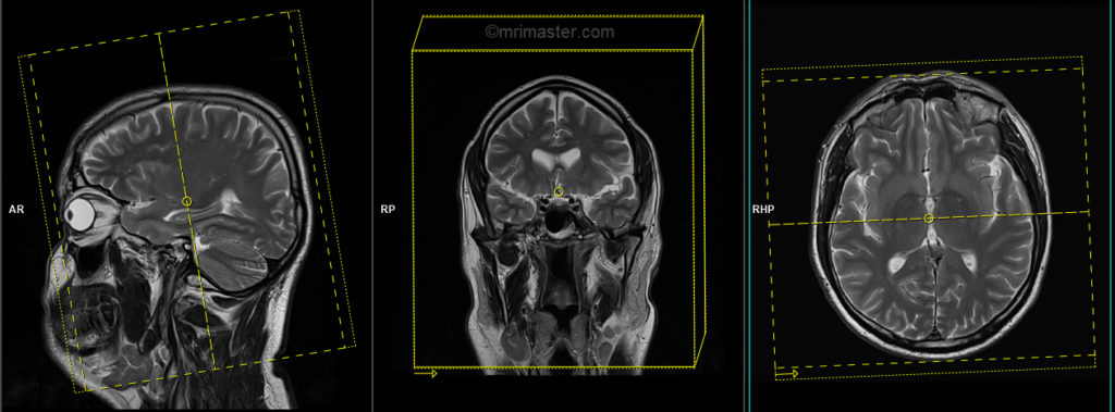
T2 tse\FLAIR axial oblique Hippocampus 2mm
Plan the axial high-resolution slices on the sagittal plane and angle the planning block parallel to the long axis of the hippocampus. Check the planning block in the other two planes. Provide an appropriate angle in the coronal plane, which should be perpendicular to the midline of the brain. Ensure that the slices are sufficient to cover the entire temporal lobe.
T2 tse\FLAIR sagittal Hippocampus 2mm SFOV
Plan the sagittal slices on the axial plane and align the block parallel to the brain’s midline. Confirm the planning block in the remaining two planes. Adjust the planning block’s angle in the coronal plane to ensure parallel alignment with the midline of the brain and the 4th ventricle. Ensure an adequate number of slices to cover the entire temporal lobe, spanning from the right side to the left side. Keep the field of view (FOV) small, typically around 120mm, to accommodate the temporal lobe of the brain.