Face MRI
Indications for face MRI scan
- Infections of bone, joint or soft tissue (eg. osteomyelitis )
- Neoplasms of bones and soft tissues
- Staging of known malignancy of face
- Treatment planning for radiation therapy
- Evaluation of response to treatment
- Pre-operative evaluation of tumours
- Presence of a foreign body
- Facial abscess
- Parotid tumour
- Orbital tumours
- Trauma
Contraindications
- Any electrically, magnetically or mechanically activated implant (e.g. cardiac pacemaker, insulin pump biostimulator, neurostimulator, cochlear implant, and hearing aids)
- Intracranial aneurysm clips (unless made of titanium)
- Pregnancy (risk vs benefit ratio to be assessed)
- Ferromagnetic surgical clips or staples
- Metallic foreign body in the eye
- Metal shrapnel or bullet
Patient preparation face MRI scan
- A satisfactory written consent form must be taken from the patient before entering the scanner room
- Ask the patient to remove all metal objects including keys, coins, wallet, cards with magnetic strips, jewellery, hearing aid and hairpins
- If possible provide a chaperone for claustrophobic patients (e.g. relative or staff )
- Contrast injection risk and benefits must be explained to the patient before the scan
- Gadolinium should only be given to the patient if GFR is > 30
- Offer earplugs or headphones, possibly with music for extra comfort
- Explain the procedure to the patient
- Instruct the patient to keep still
- Note the weight of the patient
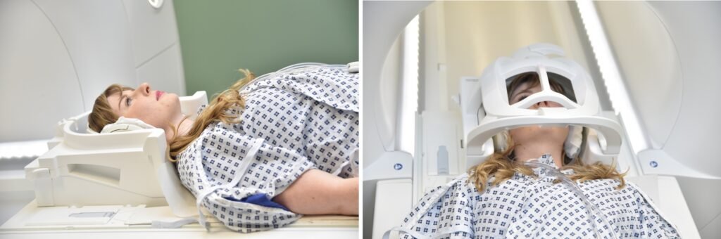
Positioning for face MRI scan
- Head first supine
- Position the head in the head coil and immobilise with cushions
- Give cushions under the legs for extra comfort
- Centre the laser beam localiser over the nose tip
Recommended Face MRI Protocols, Parameters, and Planning
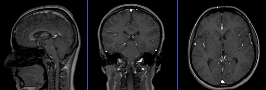
localiser Face MRI
A three-plane localizer must be taken to plan the sequences. Localizers are normally less than 25 seconds and consist of T1-weighted low-resolution scans.
T2 STIR coronal 3mm SFOV
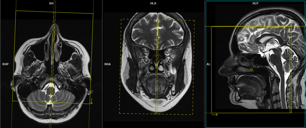
Plan the coronal slices on the sagittal plane; angle the positioning block perpendicular to the hard palate. Check the positioning block in the other two planes. An appropriate angle must be given in the axial plane (perpendicular to the nasal septum). Slices must be sufficient to cover the entire face from the tip of the nose up to the level of the fourth ventricle.
Parameters
TR 4000-5000 | TE 110 | FLIP 150 | NEX 3 | SLICE 3 MM | MATRIX 256X256 | FOV 170-180 | PHASE R>L | GAP 10% | TI 150 |
T1 tse coronal 3mm small FOV
Plan the coronal slices on the sagittal plane; angle the positioning block perpendicular to the hard palate. Check the positioning block in the other two planes. An appropriate angle must be given in the axial plane (perpendicular to the nasal septum). Slices must be sufficient to cover the entire face from the tip of the nose up to the level of the fourth ventricle.
Parameters
TR 400-600 | TE 15-25 | SLICE 3 MM | FLIP 150 | PHASE R>L | MATRIX 256X256 | FOV 170-180 | GAP 10% | NEX(AVRAGE) 2 |
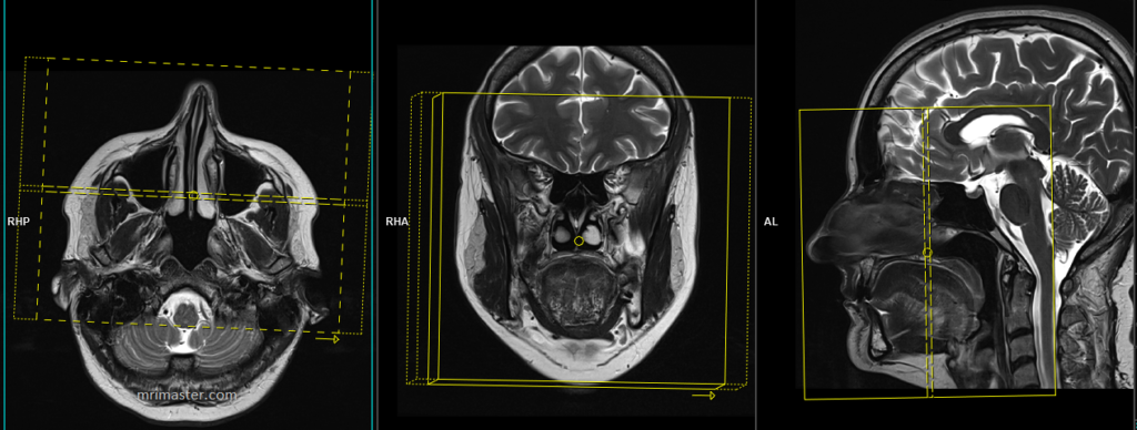
T1 tse axial 3mm SFOV
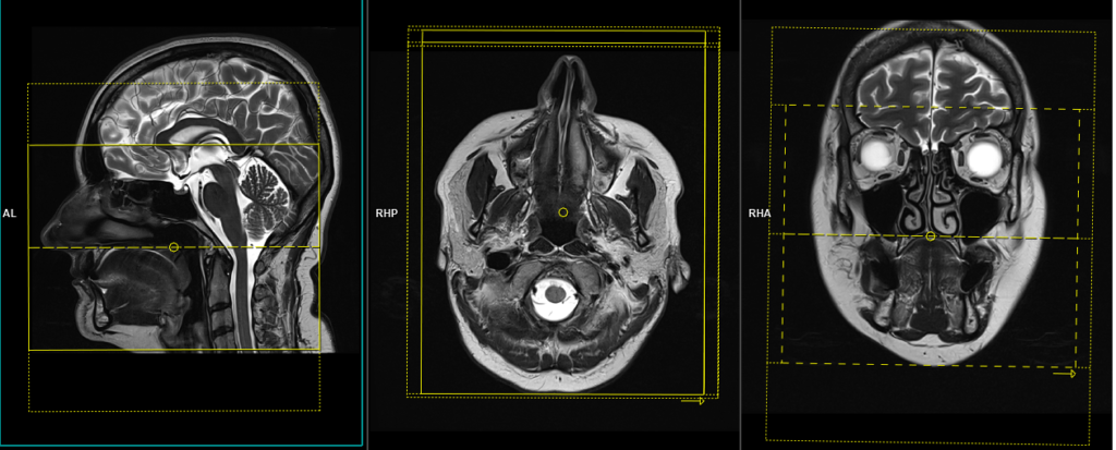
Plan the axial slices on the sagittal plane; angle the position block parallel to the hard palate. Check the positioning block in the other two planes. An appropriate angle must be given in the coronal plane (perpendicular to nasal septum). Slices must be sufficient to cover the face from the glabella down to the larynx.
Parameters
TR 400-600 | TE 15-25 | SLICE 3 MM | FLIP 140 | PHASE R>L | MATRIX 256X256 | FOV 170-180 | GAP 10% | NEX(AVRAGE) 2 |
T2 stir axial 3mm SFOV
Plan the axial slices on the sagittal plane; angle the position block parallel to the hard palate. Check the positioning block in the other two planes. An appropriate angle must be given in the coronal plane (perpendicular to nasal septum). Slices must be sufficient to cover the face from the glabella down to the larynx.

Parameters
TR 4000-5000 | TE 110 | FLIP 130 | NEX 3 | SLICE 3 MM | MATRIX 256X256 | FOV 170-180 | PHASE R>L | GAP 10% | TI 130 |
RESOLVE DWI axial 3mm
Plan the axial slices on the sagittal plane; angle the position block parallel to the hard palate. Check the positioning block in the other two planes. An appropriate angle must be given in the coronal plane (perpendicular to nasal septum). Slices must be sufficient to cover the face from the glabella down to the larynx.
Parameters
TR 7000-9000 | TE 70 | FLIP 130 | NXA 1 2 | SLICE 3MM | MATRIX 192X192 | FOV 200-230 | PHASE R>L | GAP 10% | B VALUE 0 |
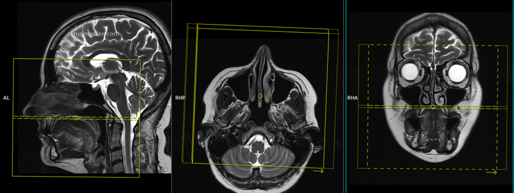
T2 tse sagittal 3mm SFOV
Plan the sagittal slices on the axial plane and angle the positioning block parallel to the nasal septum. Check the positioning block in the other two planes. An appropriate angle parallel to the nasal septum must be given in the coronal plane. The slices should be sufficient to cover the face from the right pinna to the left pinna.
Parameters
TR 3000-4000 | TE 100 | FLIP 130 | NEX 2 | SLICE 3 MM | MATRIX 256X256 | FOV 170-180 | PHASE R>L | GAP 10% | oversample 40% |
T1 VIBE DIXON 3D axial .7 mm isotropic SFOV post contrast
Plan the axial slices on the sagittal plane; angle the positioning block parallel to the hard palate. Check the positioning block in the other two planes. An appropriate angle must be given in the coronal plane (perpendicular to the nasal septum). Slices must be sufficient to cover the face from the glabella down to the larynx. Slice and phase oversampling must be given to avoid wrap-around artifacts.
TR 6.9 | TE 2.39 4.77 | FLIP 12 | NEX 2 | SLICE .7 MM | MATRIX 256X256 | FOV 170-180 | PHASE R>L | SLICES 200 | OVERSAMPLE 10% and100% |
Some radiologists prefer to perform TSE 2D scans. In that instance, use T1 TSE fat-sat small FOV axial and coronal scans after the administration of IV gadolinium DTPA injection (copy the planning outlined above).
Optional Scans
T2 space stir 3D axial 1mm isotropic SFOV
Plan the axial slices on the sagittal plane; angle the positioning block parallel to the hard palate. Check the positioning block in the other two planes. An appropriate angle must be given in the coronal plane (perpendicular to the nasal septum). Slices must be sufficient to cover the face from the glabella down to the larynx. Slice and phase oversampling must be given to avoid wrap-around artifacts.
Parameters
TR 2000-2600 | TE 200-250 | FLIP 35 | NEX 1.5 | SLICE 1 MM | MATRIX 256X256 | FOV 180-200 | PHASE R>L | GAP 10% | TI 130 |

