MRI Female Urethra
Indications for MRI urethra scan
- Assessment of pelvic floor dysfunction and pelvic organ prolapse
- Diagnosis of gonococcal and nongonococcal urethritis
- Evaluation of malignant tumours of the female urethra
- Diagnosis of cysts or abscesses of skenes gland
- Diagnosis of postirradiation injuries (e.g. urethritis)
- Evaluation of benign tumours of the urethra
- Diagnosis of cysts of bartholins gland
- Diagnosis of cysts of gartners duct
- Diagnosis of cysts of bartholins gland
- Assessment of collagen injections
- Diagnosis of urethral diverticula
- Diagnosis of tuberculosis
Contraindications
- Any electrically, magnetically or mechanically activated implant (e.g. cardiac pacemaker, insulin pump biostimulator, neurostimulator, cochlear implant, and hearing aids)
- Intracranial aneurysm clips (unless made of titanium)
- Pregnancy (risk vs benefit ratio to be assessed)
- Ferromagnetic surgical clips or staples
- Metallic foreign body in the eye
- Metal shrapnel or bullet
Patient preparation for MRI Female Urethra scan
- A satisfactory written consent form must be taken from the patient before entering the scanner room
- Ask the patient to remove all metal objects including keys, coins, wallet, cards with magnetic strips, jewellery, hearing aid and hairpins
- If possible provide a chaperone for claustrophobic patients (e.g. relative or staff )
- Contrast injection risk and benefits must be explained to the patient before the scan
- Gadolinium should only be given to the patient if GFR is > 30
- Offer earplugs or headphones, possibly with music for extra comfort
- Explain the procedure to the patient
- Instruct the patient to keep still
- Note the weight of the patient
Positioning for MRI Female Urethra scan
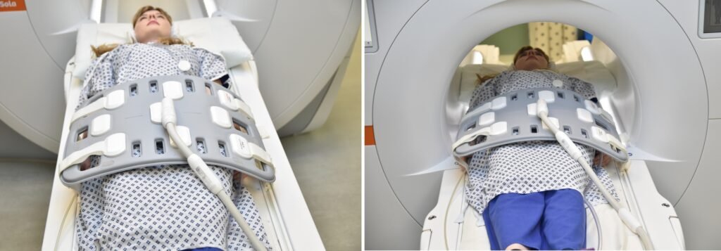
- Position the patient in supine position with head pointing towards the magnet (head first supine)
- Position the patient over the spine coil and place the body coil over the pelvis ( iliac crest down to mid thigh)
- Securely tighten the body coil using straps to prevent respiratory artefacts
- Give a pillow under the head for extra comfort
- Centre the laser beam localiser over symphysis pubis (4 inches below iliac crest)
Recommended Female Urethra MRI Protocols and Planning
MRI Female Urethra scan localiser
Take a three-plane localizer at the beginning to localize and plan the sequences. Localizers are usually completed in less than 25 seconds, and they are T1/T2-weighted low-resolution scans. Take additional localizers if needed.
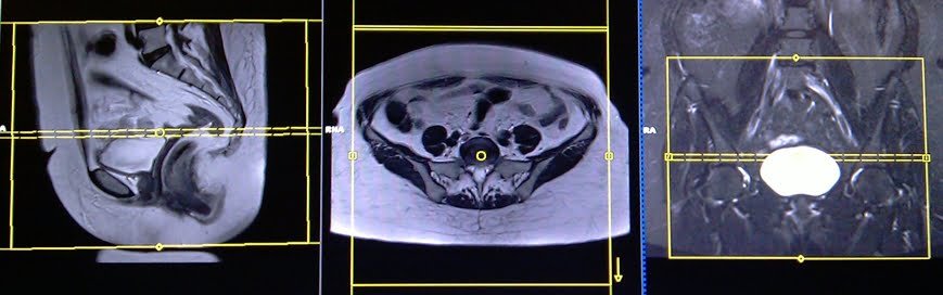
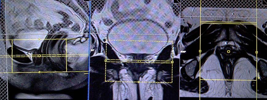
T2 tse axial 5 mm big fov
Plan the large FOV axial slices on the coronal plane; angle the positioning block parallel to the line along the right and left hip joint. Verify the positioning block in the other two planes. Establish an appropriate angle horizontally across the pelvis in the sagittal plane. The slices should be sufficient to cover the entire pelvis from the iliac crest down to two slices below the symphysis pubis. The FOV should be large enough to cover the whole pelvis (normally 350mm-400mm). Additionally, adding saturation bands on top of the axial block will reduce artefacts from arterial pulsation and breathing.
Parameters
TR 4000-5000 | TE 100-120 | SLICE 5 MM | FLIP 130-150 | PHASE A>P | MATRIX 384X384 | FOV 350-400 | GAP 10% | NEX(AVRAGE) 2 |
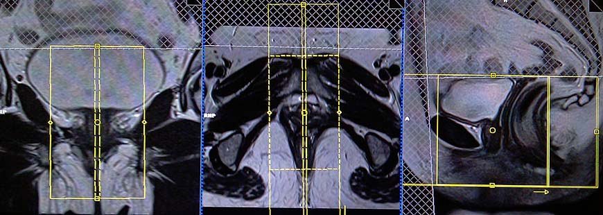
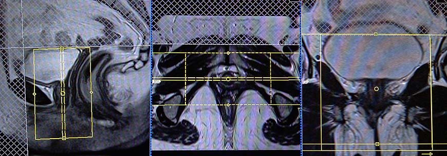
T2 tse fat sat (or stir) sagittal 3mm small fov
Plan the sagittal slices on the coronal plane; angle the positioning block parallel to the urethra (i.e., parallel to the interpubic fibrocartilage). Plan the sagittal slices on the coronal plane, positioning the block parallel to the urethra (i.e., parallel to the interpubic fibrocartilage). Verify the positioning block in the other two planes. Establish an appropriate angle in the axial plane (parallel to the interpubic fibrocartilage to the anal canal). Ensure that the slices cover the entire urethra from right to left. Use a smaller FOV around 130-150mm. To reduce artefacts from arterial pulsation, peristalsis, and breathing, add saturation bands on top and in front of the sagittal block.
Parameters
TR 5000-6000 | TE 110 | FLIP 160 | NEX 5 | SLICE 3MM | MATRIX 224X208 | FOV 130-150 | PHASE A>P | GAP 10% | FATSAT SPAIR |
T2 tse sagittal 3mm small fov
Plan the sagittal slices on the coronal plane; angle the positioning block parallel to the urethra (i.e., parallel to the interpubic fibrocartilage). Plan the sagittal slices on the coronal plane, positioning the block parallel to the urethra (i.e., parallel to the interpubic fibrocartilage). Verify the positioning block in the other two planes. Establish an appropriate angle in the axial plane (parallel to the interpubic fibrocartilage to the anal canal). Ensure that the slices cover the entire urethra from right to left. Use a smaller FOV around 130-150mm. To reduce artefacts from arterial pulsation, peristalsis, and breathing, add saturation bands on top and in front of the sagittal block.
Parameters
TR 3000-4000 | TE 110 | FLIP 160 | NEX 4 | SLICE 3MM | MATRIX 256X224 | FOV 120-150 | PHASE A>P | GAP 10% | FATSAT OFF |
T2 tse fat sat (or stir) axial OBLIQUE 3mm small fov
Plan the axial slices on the sagittal plane; angle the positioning block perpendicular to the urethra. Check the positioning block in the other two planes. An appropriate angle must be given in the coronal plane (perpendicular to the urethra). The slices must be sufficient to cover the whole urethra from the mid-urinary bladder down to 1 inch below the opening of the urethra. Use a smaller FOV around 130-150mm. Adding saturation bands on top and in front of the axial block will reduce artifacts from arterial pulsation, peristalsis, and breathing.
Parameters
TR 4000-5000 | TE 110 | FLIP 160 | NEX 4 | SLICE 3MM | MATRIX 224X208 | PHASE A>P | GAP 10% | FATSAT SPAIR |
T2 tse axial oblique 3mm small fov
Plan the axial slices on the sagittal plane; angle the positioning block perpendicular to the urethra. Check the positioning block in the other two planes. An appropriate angle must be given in the coronal plane (perpendicular to the urethra). The slices must be sufficient to cover the whole urethra from the mid-urinary bladder down to 1 inch below the opening of the urethra. Use a smaller FOV around 130-150mm. Adding saturation bands on top and in front of the axial block will reduce artifacts from arterial pulsation, peristalsis, and breathing.
Parameters
TR 3000-4000 | TE 110 | FLIP 160 | NEX 4 | SLICE 3MM | MATRIX 256X224 | PHASE A>P | GAP 10% | FATSAT OFF |
T1 tse fat sat axial oblique 3mm small fov
Plan the axial slices on the sagittal plane; angle the positioning block perpendicular to the urethra. Check the positioning block in the other two planes. An appropriate angle must be given in the coronal plane (perpendicular to the urethra). The slices must be sufficient to cover the whole urethra from the mid-urinary bladder down to 1 inch below the opening of the urethra. Use a smaller FOV around 130-150mm. Adding saturation bands on top and in front of the axial block will reduce artifacts from arterial pulsation, peristalsis, and breathing.
Parameters
TR 400-500 | TE 15-20 | FLIP 130 | NEX 5 | SLICE 3MM | MATRIX 224X224 | PHASE A>P | GAP 10% | FATSAT ON |
T2 tse fat sat (or stir)coronal oblique 3mm
Plan the coronal slices on the sagittal plane; angle the positioning block parallel to the urethra. Check the positioning block in the other two planes. An appropriate angle must be given in the axial plane (parallel to the right and left ischial tuberosities). The slices must be sufficient to cover the whole urethra from mid symphysis pubis to mid rectum. Adding saturation bands on top and in front of the coronal block will reduce artifacts from arterial pulsation, peristalsis, and breathing.
Parameters
TR 4000-5000 | TE 110 | FLIP 150 | NEX 2 | SLICE 3 MM | MATRIX 224X224 | PHASE R>L | GAP 10% | FAT SAT SPAIR |
Very rarely some urethra scans needed contrast enhanced imaging. In case of contrast enhanced imaging use T1 TSE fat saturated small FOV axial and coronal obliques after the administration of IV gadolinium DTPA injection(copy the planning outlined above). The document below provides access to the recommended dosage of gadolinium DTPA injection, as advised by the manufacturer.
CLICK THE SEQUENCES BELOW TO CHECK THE SCANS
- localizer_3 plane1
- T2_TSE_AXIAL BIG FOV_5MM2
- T2_TSE_SAGITTAL_SFOV_3MM3
- T2_TSE_FAT SAT_SAGITTAL_SFOV_3MM4
- T2_TSE_FAT SAT_AXIAL_OBLIQUE_SFOV_3MM5
- T2_TSE_AXIAL OBLIQUE_SFOV_3MM6
- T1_TSE_FAT SAT_AXIAL OBLIQUE_SFOV_3MM7
- T2_TSE FAT SAT_CORONAL_OBLIQUE_SFOV_3MM8
- CONTRAST ENHANCEMENT

- T1_TSE_FAT SAT_AXIAL OBLIQUE_SFOV_3MM POST9
- T1_TSE_FAT SAT_CORONAL OBLIQUE_SFOV_3MM POST10

