MRI Upper Arm (Humerus): Protocol and Planning
Indications for MRI Upper Arm (Humerus)
- Marrow abnormalities (e.g. bone contusions, osteonecrosis, marrow oedema syndromes, and stress fractures)
- Synovial-based disorders ( e.g. synovitis, tenosynovitis, bursitis, and ganglion cysts)
- Infections of bone, joint, or soft tissue (e.g. osteomyelitis, osteochondritis, osteoarthritis )
- Evaluation of suspected cartilaginous defects
- Neoplasms of bone, joint or soft tissue
- Avascular necrosis
- Nerve Impingement
- Fractures in children
- Soft-tissue masses
- Occult fracture
- Ligament tear
Contraindications
- Any electrically, magnetically or mechanically activated implant (e.g. cardiac pacemaker, insulin pump biostimulator, neurostimulator, cochlear implant, and hearing aids)
- Intracranial aneurysm clips (unless made of titanium)
- Pregnancy (risk vs benefit ratio to be assessed)
- Ferromagnetic surgical clips or staples
- Metallic foreign body in the eye
- Metal shrapnel or bullet
Patient preparation for MRI Upper Arm (Humerus)
- A satisfactory written consent form must be taken from the patient before entering the scanner room
- Ask the patient to remove all metal objects including keys, coins, wallet, cards with magnetic strips, jewellery, hearing aid and hairpins
- If possible provide a chaperone for claustrophobic patients (e.g. relative or staff )
- Offer earplugs or headphones, possibly with music for extra comfort
- Explain the procedure to the patient
- Instruct the patient to keep still
- Note the weight of the patient
Positioning for MRI Upper Arm (Humerus)
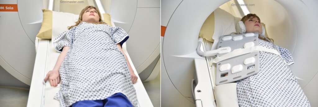
- Position the patient in supine position with head pointing towards the magnet (head first supine)
- Position the patient off-center over the spine coil, as demonstrated. Place the body coil over the upper arm, from the shoulder to the elbow.
- Securely tighten the body coil using straps to prevent respiratory artifacts
- Give a pillow under the head and cushions under the legs for extra comfort
- Centre the laser beam localiser over the mid humerus.
- Register the patient on the scanner as 'head first supine'
Recommended MRI Upper Arm (Humerus) Protocols and Planning
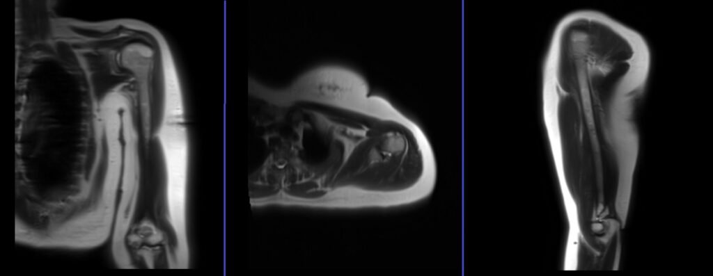
localiser
A three-plane localizer must be taken at the beginning to localize and plan the sequences. Localizers are usually less than 25 seconds. T1\T2-weighted low-resolution scans can be used for this purpose. Continue taking additional localizers until a true coronal, axial, and sagittal view of the upper arm is obtained.
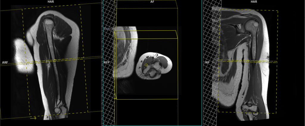
T2 stir axial 3mm SFOV
Plan the axial slices on the coronal plane and angle the planning block perpendicular to the humerus. Check the planning block in the other two planes. An appropriate angle must be used in the sagittal plane (perpendicular to the humerus). Slices must be sufficient to cover the whole upper arm from the acromioclavicular joint to the elbow joint. Adding saturation bands over the chest will help reduce breathing artifacts. The phase direction must be anteroposterior to avoid wrap-around and motion artifacts from the chest.
Parameters
TR 4000-6000 | TE 110 | FLIP 160 | NEX 2 | SLICE 6 MM | MATRIX 256X256 | FOV 170-200 | PHASE A>P | GAP 10% | TI 150 |
T1 tse axial 3mm SFOV
Plan the axial slices on the coronal plane and angle the planning block perpendicular to the humerus. Check the planning block in the other two planes. An appropriate angle must be used in the sagittal plane (perpendicular to the humerus). Slices must be sufficient to cover the whole upper arm from the acromioclavicular joint to the elbow joint. Adding saturation bands over the chest will help reduce breathing artifacts. The phase direction must be anteroposterior to avoid wrap-around and motion artifacts from the chest.
Parameters
TR 400-600 | TE 15-25 | SLICE 6 MM | FLIP 160 | PHASE A>P | MATRIX 320X320 | FOV 170-200 | GAP 10% | NEX(AVRAGE) 2 |
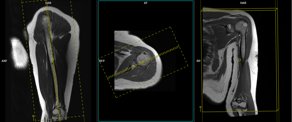
T1 tse coronal 3mm
Plan the coronal slices on the axial plane and angle the planning block parallel to the scapular blade. Check the planning block in the other two planes. An appropriate angle must be used in the sagittal plane (parallel to the humerus). Slices must be sufficient to cover the whole upper arm from anterior to posterior. The field of view (FOV) must be big enough to cover both the shoulder and elbow joints. Adding saturation bands over the chest will reduce breathing and arterial pulsation artifacts. For the phase direction, you can choose either right to left or head to feet, and a minimum 100% oversampling is recommended to avoid wrap-around artifacts.
Parameters
TR 400-600 | TE 15-25 | SLICE 5 MM | FLIP 150 | PHASE R>L | MATRIX 448X384 | FOV 350-400 | GAP 10% | NEX(AVRAGE) 2 |
T2 stir coronal 3mm
Plan the coronal slices on the axial plane and angle the planning block parallel to the scapular blade. Check the planning block in the other two planes. An appropriate angle must be used in the sagittal plane (parallel to the humerus). Slices must be sufficient to cover the whole upper arm from anterior to posterior. The field of view (FOV) must be big enough to cover both the shoulder and elbow joints. Adding saturation bands over the chest will reduce breathing and arterial pulsation artifacts. For the phase direction, you can choose either right to left or head to feet, and a minimum 100% oversampling is recommended to avoid wrap-around artifacts.
Parameters
TR 3000-4000 | TE 110 | FLIP 150 | NEX 2 | SLICE 5 MM | MATRIX 384X320 | FOV 350-400 | PHASE R>L | GAP 10% | TI 150 |
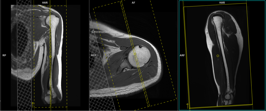
T2 tse sagittal 3mm
Plan the sagittal slices on the axial plane and angle the planning block perpendicular to the scapular blade. Check the planning block in the other two planes. Use an appropriate angle in the coronal plane (parallel to the humerus). The slices must sufficiently cover the entire upper arm from the medial to lateral aspect. The field of view (FOV) should be large enough to encompass both the shoulder and elbow joints. To reduce artifacts from breathing and arterial pulsations, consider adding saturation bands over the chest. For the phase direction, you can choose either anterior to posterior or head to feet. A minimum oversampling of 100% is recommended to avoid wrap-around artifacts.
Parameters
TR 4000-5000 | TE 110 | FLIP 130 | NEX 2 | SLICE 5MM | MATRIX 384X384 | FOV 350-400 | PHASE A>P | GAP 10% | FATSAT Off |

