MRI Kidney Ureters and Bladder (KUB) Protocols and Planning
Indications for mri kidney ureters and bladder (KUB) scan
- Detection of suspected stones where CT is contraindicated (post radiotherapy , children )
- For the evaluation of solid abdominal masses
- For the evaluation of malignancy
- For the evaluation of cyst
- Local staging of cancer
- Genitourinary tract TB
- Haematuria
- Trauma
Contraindications for MRI Kidney Ureters and Bladde Scan
- Any electrically, magnetically or mechanically activated implant (e.g. cardiac pacemaker, insulin pump biostimulator, neurostimulator, cochlear implant, and hearing aids)
- Intracranial aneurysm clips (unless made of titanium)
- Pregnancy (risk vs benefit ratio to be assessed)
- Ferromagnetic surgical clips or staples
- Metallic foreign body in the eye
- Metal shrapnel or bullet
Patient preparation MRI Kidney Ureters and Bladder (KUB) scan
- A satisfactory written consent form must be taken from the patient before entering the scanner room
- Ask the patient to remove all metal object including keys, coins, wallet, any cards with magnetic strips, jewellery, hearing aid and hairpins
- Ask the patient to undress and change into a hospital gown
- Claustrophobic patients may be accompanied into the scanner room e.g. by staff member or relative with proper safety screening
- Offer headphones for communicating with the patient and ear protection
- Explain the procedure to the patient and answer questions
- Note the weight of the patient
- Patient should be have a full balder
Positioning MRI Kidney Ureters and Bladder (KUB)
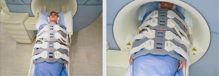
- Position the patient in supine position with head pointing towards the magnet (head first supine)
- Position the patient over the spine coil and place the body coils over abdomen and pelvis (nipple down to elbow three inches below symphysis pubis)
- Securely tighten the body coil using straps to prevent respiratory artefacts
- Give a pillow under the head and cushions under the legs for extra comfort
- Centre the laser beam localiser over the iliac crest
- Register the patient in the scanner as head first supine
Recommended MRI Kidney Ureters and Bladder (KUB) Protocols and Planning
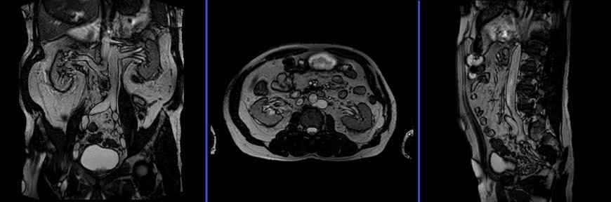
MRI Kidney Ureters and Bladder (KUB) Localizer
A three-plane T2 TRIFISP\HASTE localizer must be taken initially to localize and plan the sequences. These are fast single-shot localizers with under 25s acquisition time, which are excellent for localizing vascular structures. Take at least 5-8 slices in all planes to get the best results.
T2 HASTE\TRUFI axial 4 mm
Plan the axial slices on the coronal plane, angle the positioning block parallel to the right and left iliac crest. Check the positioning block in the other two planes. An appropriate angle must be given in the sagittal plane (horizontally across the abdomen). The slices must be sufficient to cover the entire abdomen and pelvis from the diaphragm down to the symphysis pubis. Use a field of view (FOV) that is large enough to encompass the entire abdomen, typically ranging from 350mm to 400mm.
Parameters
TR 4-5 | TE 2-3 | FLIP 60 | NEX 1 | SLICE 4 MM | MATRIX 320×256 | FOV 350-400 | PHASE R>L | OVERSAMPLE 50% | IPAT ON |
T2 HASTE\ TRUFI fat sat axial 4 mm
Plan the axial slices on the coronal plane, angle the positioning block parallel to the right and left iliac crest. Check the positioning block in the other two planes. An appropriate angle must be given in the sagittal plane (horizontally across the abdomen). The slices must be sufficient to cover the entire abdomen and pelvis from the diaphragm down to the symphysis pubis. Use a field of view (FOV) that is large enough to encompass the entire abdomen, typically ranging from 350mm to 400mm.
Parameters
TR 4-5 | TE 2-3 | FLIP 60 | NEX 1 | SLICE 4 MM | MATRIX 320×256 | FOV 350-400 | PHASE R>L | OVERSAMPLE 50% | fat sat ON |
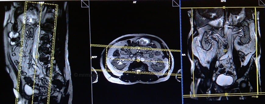
T1 vibe DIXON 3d axial 4 mm
Plan the axial slices on the coronal plane; angle the position block parallel to the line along the right and left iliac crest. Check the positioning block in the other two planes. An appropriate angle must be given in the sagittal plane (perpendicular to the lumbar spine). Slices must be sufficient to cover the whole abdomen and pelvis from diaphragm down to symphysis pubis. FOV must be big enough to cover the whole abdomen (normally 350mm-400mm).
Parameters
TR 6-7 | TE 2.39 4.77 | FLIP 10 | NXA 1 | SLICE 4 MM | MATRIX 320×320 | FOV 320-350 | PHASE A>P | OVERSAMPLE 20% | BH YES |
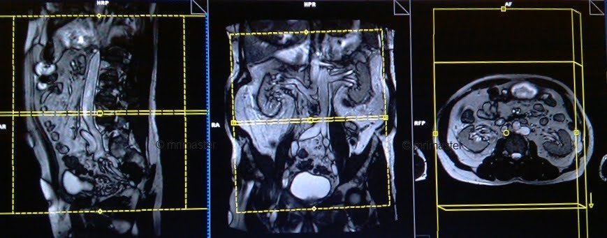
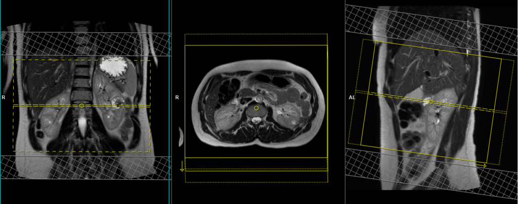
T1 vibe DIXON 3D coronal 2mm
Plan the coronal slices on the axial plane; angle the position block parallel to the mid line along the right and left kidneys. Check the positioning block in the other two planes. An appropriate angle must be given in the sagittal plane (parallel to L spine). Slices must be sufficient to cover the whole urinary system. Phase oversampling and, in the case of 3D blocks, slice oversample, must be used to avoid wrap around artefacts. FOV must be big enough to cover the kidneys and bladder. Instruct the patient to hold their breath during image acquisition. (In our department we instruct the patients to breath in and out twice before the “breath in and hold” instruction.).
Parameters
TR 6-7 | TE 2.39 4.77 | FLIP 10 | NXA 1 | SLICE 2 MM | MATRIX 320×320 | FOV 320-350 | PHASE R>L | OVERSAMPLE 20% | BH YES |
T2 HASTE fat sat coronal 3mm
Plan the coronal slices on the axial plane; angle the position block parallel to the mid line along the right and left kidneys. Check the positioning block in the other two planes. An appropriate angle must be given in the sagittal plane (parallel to L spine). Slices must be sufficient to cover the whole urinary system. Phase oversampling and, in the case of 3D blocks, slice oversample, must be used to avoid wrap around artefacts. FOV must be big enough to cover the kidneys and bladder. Instruct the patient to hold their breath during image acquisition. (In our department we instruct the patients to breath in and out twice before the “breath in and hold” instruction.).
Parameters
TR 2000-2500 | TE 90-100 | FLIP 150 | NEX 1 | SLICE 3 MM | MATRIX 320X320 | FOV 400-450 | PHASE R>L | OVERSAMPLE 50% | IPAT OFF |
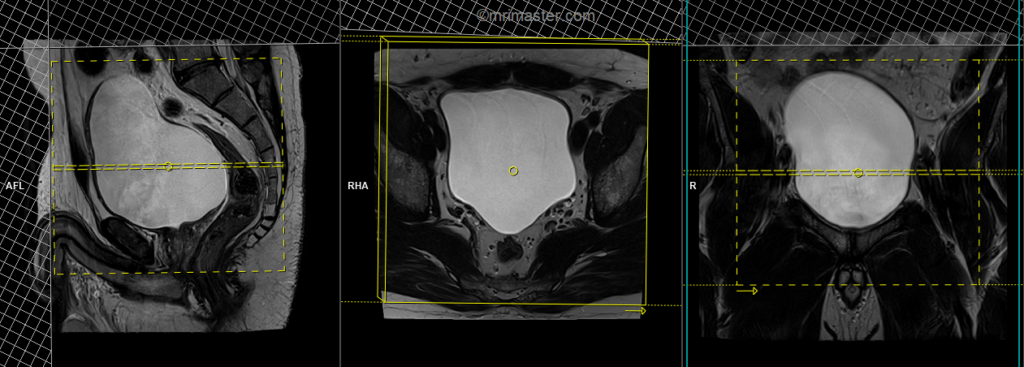
T2 tse axial 3mm SFOV
Plan the axial slices on the sagittal plane and angle the positioning block horizontally across the urinary bladder. Check the positioning block in the other two planes. An appropriate angle must be given in the coronal plane, horizontally across the urinary bladder. Slices should be sufficient to cover the urinary bladder from L1 down to two slices below the symphysis pubis. To reduce artifacts from breathing and peristalsis, consider using a saturation band above and in front of the axial block.
Parameters
TR 4000-6000 | TE 100-120 | SLICE 3 MM | FLIP 130-150 | PHASE R>L | MATRIX 320X320 | FOV 200-250 | GAP 10% | NEX(AVRAGE) 2 |
T2 tse\haste fat sat breath hold 4mm axial
Plan the coronal slices on the axial plane and angle the positioning block parallel to the right and left kidneys. Check the positioning block in the other two planes. Ensure an appropriate angle is given in the sagittal plane (parallel to the long axis of the kidney). The slices should adequately cover both kidneys from the anterior to posterior direction. Phase oversampling should be used to prevent wrap-around artifacts. Consider adding saturation bands at the top and bottom of the block to minimize artifacts caused by fat signal, arterial pulsation, and breathing. Instruct the patient to hold their breath during image acquisition.
Parameters HASTE FAT SAT
TR 2000-2500 | TE 90-110 | FAT SAT SPAIR | NEX 1 | SLICE 4MM | MATRIX 320×256 | FOV 300 | PHASE R>L | OVERSAMPLE 50% | TRIGGER NO |
Parameters T2 FAT SAT
TR 6000-7000 | TE 150 | FAT SAT SPAIR | NEX 1 | SLICE 4MM | MATRIX 256×208 | FOV 280-300 | PHASE A>P | OVERSAMPLE 80% | IPAT ON |

