LOWER LEg
Indications mri lower leg (tibia and fibula)
- Marrow abnormalities (eg. bone contusions, osteonecrosis, marrow edema syndromes, and stress fractures)
- Infections of bone, joint, or soft tissue (eg. osteomyelitis , osteochondritis ,osteo arthritis )
- Neoplasms of bone, joint, or soft tissue
- Avascular necrosis
- Nerve impingement
- Fractures in children
- Soft-tissue masses
- Occult fracture
- Ligament tear
- Cysts
Contraindications
- Any electrically, magnetically or mechanically activated implant (e.g. cardiac pacemaker, insulin pump biostimulator, neurostimulator, cochlear implant, and hearing aids)
- Intracranial aneurysm clips (unless made of titanium)
- Pregnancy (risk vs benefit ratio to be assessed)
- Ferromagnetic surgical clips or staples
- Metallic foreign body in the eye
- Metal shrapnel or bullet
Patient preparation
- A satisfactory written consent form must be taken from the patient before entering the scanner room
- Ask the patient to remove all metal object including keys, coins, wallet, any cards with magnetic strips, jewellery, hearing aid and hairpins
- If possible provide a chaperone for claustrophobic patients (e.g. relative or staff )
- Offer earplugs or headphones, possibly with music for extra comfort
- Explain the procedure to the patient
- Instruct the patient to keep still
- Note the weight of the patient
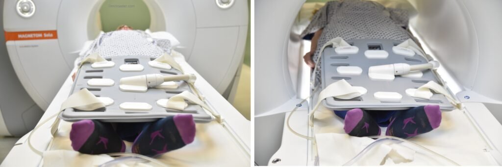
Positioning
- Place the patient in a supine position, either with their feet pointing towards the magnet (feet first supine) or with their head pointing towards the magnet (head first supine).
- Position the patient over the spine coil and place the body coils over the lower leg (knee joint down to ankle joint)
- Securely tighten the body coil using straps
- Give a pillow under the head for extra comfort
- Centre the laser beam localiser over the middle of lower leg
Suggested sequence, parameters and planning
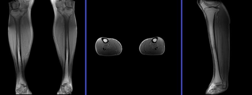
localiser
A three-plane localizer must be taken at the beginning to localize and plan the sequences. Localizers are normally less than 25 seconds and are T1-weighted low-resolution scans.
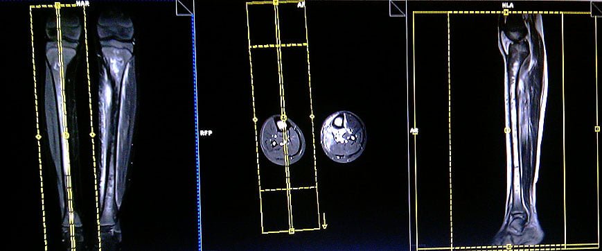
T2 stir coronal 4mm 460 FOV
Plan the coronal slices on the sagittal plane and angle the positioning block parallel to the tibial shaft. Verify the positioning block in the other two planes. Ensure an appropriate angle is set in the axial plane, aligned with the right and left shafts of the tibia. The slices should adequately cover the entire lower leg from anterior to posterior. The field of view (FOV) should be large enough to encompass both the knee joint and ankle joint (typically ranging from 450mm to 480mm).
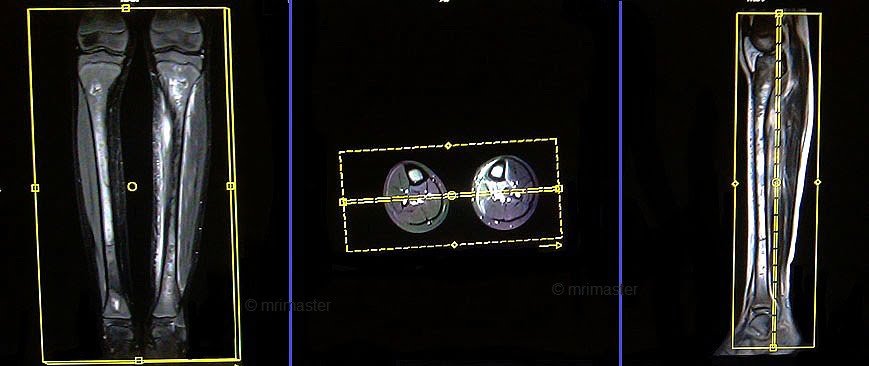
Parameters
TR 4000-5000 | TE 110 | FLIP 160 | NEX 2 | SLICE 3 MM | MATRIX 448X384 | FOV 450-480 | PHASE R>L | GAP 10% | TI 150 |
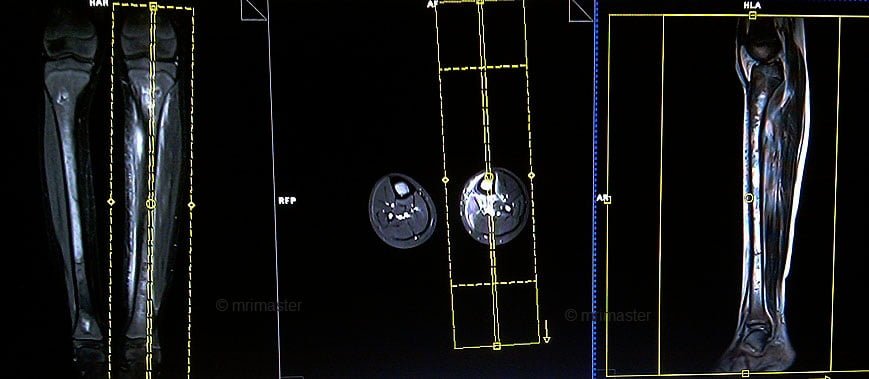
T1 tse coronal 4mm 460 FOV
Plan the coronal slices on the sagittal plane and angle the positioning block parallel to the tibial shaft. Verify the positioning block in the other two planes. Ensure an appropriate angle is set in the axial plane, aligned with the right and left shafts of the tibia. The slices should adequately cover the entire lower leg from anterior to posterior. The field of view (FOV) should be large enough to encompass both the knee joint and ankle joint (typically ranging from 450mm to 480mm).
Parameters
TR 400-600 | TE 15-25 | SLICE 3 MM | FLIP 160 | PHASE A>P | MATRIX 448X448 | FOV 450-480 | GAP 10% | NEX(AVRAGE) 2 |
T1 tse axial 6mm
Plan the axial slices on the sagittal plane and angle the positioning block perpendicular to the shaft of the tibia. Verify the positioning block in the other two planes. An appropriate angle should be applied in the coronal plane, perpendicular to the right and left shaft of the tibia. Ensure that the slices cover the entire lower leg from the knee joint down to the ankle joint. In case the pathology is localized in one area, perform axial slices only in that specific region.
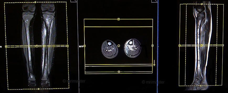
Parameters
TR 400-600 | TE 15-25 | SLICE 6MM | FLIP 160 | PHASE A>P | MATRIX 512X448 | FOV 350-400 | GAP 10% | NEX(AVRAGE) 2 |
T2 stir axial 6mm
Plan the axial slices on the sagittal plane and angle the positioning block perpendicular to the shaft of the tibia. Verify the positioning block in the other two planes. An appropriate angle should be applied in the coronal plane, perpendicular to the right and left shaft of the tibia. Ensure that the slices cover the entire lower leg from the knee joint down to the ankle joint. In case the pathology is localized in one area, perform axial slices only in that specific region.
Parameters
TR 3000-4000 | TE 110 | FLIP 160 | NEX 2 | SLICE 6MM | MATRIX 448X384 | FOV 350-400 | PHASE A>P | GAP 10% | TI 150 |
T2 tse sagittal 3mm right
Plan the right-side sagittal slices on the coronal plane and angle the positioning block parallel to the shaft of the right tibia. An appropriate angle must be given in the axial plane (vertically across the tibia). Slices must be sufficient to cover the right lower leg from medial to lateral. The field of view (FOV) must be big enough to cover the knee joint and ankle joint (normally 450mm to 480mm).
Parameters
TR 4000-5000 | TE 100 | SLICE 3 MM | FLIP 160 | PHASE A>P | MATRIX 512X448 | FOV 450-480 | GAP 10% | NEX(AVRAGE) 2 |
T2 tse sagittal 3mm left
Plan the left-side sagittal slices on the coronal plane and angle the positioning block parallel to the shaft of the left tibia. An appropriate angle must be given in the axial plane (vertically across the tibia). Slices must be sufficient to cover the left lower leg from medial to lateral. The field of view (FOV) must be big enough to cover the knee joint and ankle joint (normally 450mm to 480mm).
Parameters
TR 4000-5000 | TE 100 | SLICE 3 MM | FLIP 160 | PHASE A>P | MATRIX 512X448 | FOV 450-480 | GAP 10% | NEX(AVRAGE) 2 |

