MRI Kidneys (Renal MRI)
Indications for MRI kidney scan
- For the assessment of malignant renal lesions (e.g. renal cell carcinomas and transitional cell carcinoma).
- For the assessment of benign renal lesions (e.g. oncocytoma and angiomyolipoma)
- For the assessment of the inferior vena cava in patients with known solid renal tumour
- For the assessment of xanthogranulomatous pyelonephritis
- For the assessment of cystic kidney disease
- Hematuria
Contraindications
- Any electrically, magnetically or mechanically activated implant (e.g. cardiac pacemaker, insulin pump biostimulator, neurostimulator, cochlear implant, and hearing aids)
- Intracranial aneurysm clips (unless made of titanium)
- Pregnancy (risk vs benefit ratio to be assessed)
- Ferromagnetic surgical clips or staples
- Metallic foreign body in the eye
- Metal shrapnel or bullet
Patient preparation for MRI kidney scan
- A satisfactory written consent form must be taken from the patient before entering the scanner room
- Ask the patient to remove all metal objects including keys, coins, wallet, cards with magnetic strips, jewellery, hearing aid and hairpins
- Ask the patient to undress and change into a hospital gown
- Instruct the patient to hold their breath for the breath hold scans (its better to coach the patient two to three times before starting the scan)
- An intravenous line must be placed with extension tubing extending out of the magnetic bore
- Contrast injection risk and benefits must be explained to the patient before the scan
- Gadolinium should only be given to the patient if GFR is > 30
- If possible provide a chaperone for claustrophobic patients (e.g. relative or staff )
- Offer earplugs or headphones, possibly with music for extra comfort
- Explain the procedure to the patient
- Instruct the patient to keep still
- Note the weight of the patient
Positioning for MRI kidney scan
- Position the patient in supine position with head pointing towards the magnet (head first supine)
- Position the patient over the spine coil and place the body coil over the abdomen (xiphoid process down to anterior superior iliac spine)
- Securely tighten the body coil using straps to prevent respiratory artefacts
- Give a pillow under the head and cushions under the legs for extra comfort
- Centre the laser beam localiser over the level of lower intercostal border

Recommended MRI kidney Protocols, Parameters, and Planning
MRI Kidney localiser
To localize and plan the sequences, it is essential to acquire a three-plane T2 HASTE localizer initially. These fast single-shot localizers have an acquisition time of under 25 seconds and are highly effective in accurately localizing abdominal structures.

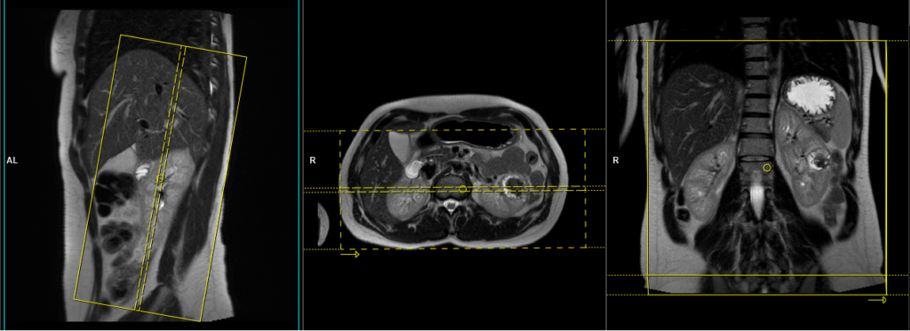
T2 HASTE\t2 tse coronal 4mm
Plan the coronal slices on the axial plane and angle the positioning block parallel to the right and left kidneys. Check the positioning block in the other two planes. Provide an appropriate angle in the sagittal plane (parallel to the long axis of the kidney). Ensure that the slices are sufficient to cover both kidneys from anterior to posterior. Phase oversampling must be used to avoid wrap-around artifacts. Instruct the patient to hold their breath during image acquisition. (In our department, we instruct patients to take two breaths in and out before giving the instruction to ‘breathe in and hold’.)
Parameters haste
TR 2000-2500 | TE 90-110 | FLIP 130 | NEX 1 | SLICE 4MM | MATRIX 320×256 | FOV 300 | PHASE R>L | OVERSAMPLE 50% | TRIGGER NO |
Parameters t2 tse
| TR 5000-6000 | TE 150 | FLIP 150 | NEX 1 | SLICE 4MM | MATRIX 256×224 | FOV 300 | PHASE R>L | OVERSAMPLE 50% | IPAT ON |
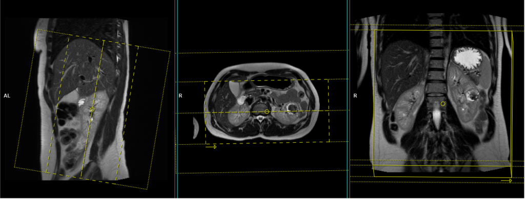
T1 vibe DIXON breath hold coronal 4mm
Plan the coronal slices on the axial plane and angle the positioning block parallel to the right and left kidneys. Check the positioning block in the other two planes. An appropriate angle must be given in the sagittal plane, parallel to the long axis of the kidneys. Ensure that the slices are sufficient to cover both kidneys from anterior to posterior. Phase oversampling and slice oversampling must be used to avoid wrap-around artifacts. Instruct the patient to hold their breath during image acquisition. In our department, we instruct patients to breathe in and out twice before giving the ‘breathe in and hold’ instruction.
Parameters VIBE dixon
TR 6-7 | TE 2.39 4.77 | FLIP 10 | NXA 1 | SLICE 4 MM | MATRIX 320×320 | FOV 320-350 | PHASE A>P | OVERSAMPLE 20% | BH YES |
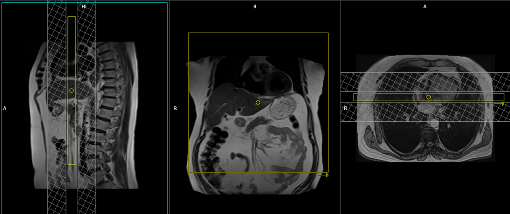
T2 tse\haste breath hold 4mm axial
Plan the axial slices on the coronal plane; angle the position block parallel to the right and left renal pelvis. Check the positioning block in the other two planes. An appropriate angle must be given in the sagittal plane (perpendicular to the long axis of kidney). Slices must be sufficient to cover both kidneys from two slices above the upper pole of kidneys down to two slices below the lower pole of kidney. Phase oversampling and, in the case of 3D blocks, slice oversample, must be used to avoid wrap around artefacts. Consider adding saturation bands at the top and bottom of the block to minimize artifacts caused by fat signal, arterial pulsation, and breathing. Instruct the patient to hold their breath during image acquisition.
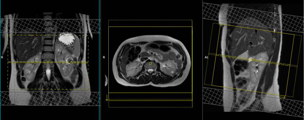
When planning the axial breath-hold scans, it is crucial to utilize the breath-hold vibe coronal sequence. This is because during inhalation, the diaphragm exerts downward pressure on the liver, causing a shift in its position from the initial localizer scans. Therefore, to accurately capture the desired imaging area of the liver, it is important to account for this positional change by utilizing the breath-hold vibe coronal sequence.

Parameters haste
TR 2000-2500 | TE 90-110 | FLIP 130 | NEX 1 | SLICE 4MM | MATRIX 320×256 | FOV 300 | PHASE R>L | OVERSAMPLE 50% | TRIGGER NO |
Parameters t2 tse
| TR 5000-6000 | TE 150 | FLIP 150 | NEX 1 | SLICE 4MM | MATRIX 256×256 | FOV 280-300 | PHASE A>P | OVERSAMPLE 80% | IPAT ON |
T2 tse\haste fat sat breath hold 4mm axial
Plan the axial slices on the coronal plane; angle the position block parallel to the right and left renal pelvis. Check the positioning block in the other two planes. An appropriate angle must be given in the sagittal plane (perpendicular to the long axis of kidney). Slices must be sufficient to cover both kidneys from two slices above the upper pole of kidneys down to two slices below the lower pole of kidney. Phase oversampling and, in the case of 3D blocks, slice oversample, must be used to avoid wrap around artefacts. Consider adding saturation bands at the top and bottom of the block to minimize artifacts caused by fat signal, arterial pulsation, and breathing. Instruct the patient to hold their breath during image acquisition.
Parameters HASTE FAT SAT
TR 2000-2500 | TE 90-110 | FAT SAT SPAIR | NEX 1 | SLICE 4MM | MATRIX 320×256 | FOV 300 | PHASE R>L | OVERSAMPLE 50% | TRIGGER NO |
Parameters T2 FAT SAT
| TR 6000-7000 | TE 150 | FAT SAT SPAIR | NEX 1 | SLICE 4MM | MATRIX 256×208 | FOV 280-300 | PHASE A>P | OVERSAMPLE 80% | IPAT ON |
DWI epi 3 scan trace axial 3mm free breathing
Plan the axial slices on the coronal free-breathing localizer; angle the position block parallel to the right and left renal pelvis. Check the positioning block in the other two planes. An appropriate angle must be given in the sagittal plane (perpendicular to the long axis of kidney). Slices must be sufficient to cover both kidneys from two slices above the upper pole of kidneys down to two slices below the lower pole of kidney. Phase oversampling and, in the case of 3D blocks, slice oversample, must be used to avoid wrap around artefacts. Consider adding saturation bands at the top and bottom of the block to minimize artifacts caused by fat signal, arterial pulsation, and breathing.
Parameters epI DWI
TR 6000-7000 | TE 90 | IPAT ON | NEX 3 5 8 | SLICE 3 MM | MATRIX 192X192 | FOV 200-250 | PHASE R>L | GAP 10% | B VALUE 0 |
T1 VIBE DIXON 3mm axial BH pre GD(In-opposed phase and water sat)
Plan the axial slices on the coronal plane; angle the position block parallel to the right and left renal pelvis. Check the positioning block in the other two planes. An appropriate angle must be given in the sagittal plane (perpendicular to the long axis of kidney). Slices must be sufficient to cover both kidneys from two slices above the upper pole of kidneys down to two slices below the lower pole of kidney. Phase oversampling and, in the case of 3D blocks, slice oversample, must be used to avoid wrap around artefacts. Consider adding saturation bands at the top and bottom of the block to minimize artifacts caused by fat signal, arterial pulsation, and breathing. Instruct the patient to hold their breath during image acquisition.
Parameters
TR 6-7 | TE 2.39 4.77 | FLIP 10 | NXA 1 | SLICE 3 MM | MATRIX 320×320 | FOV 320-350 | PHASE A>P | OVERSAMPLE 20% | BH YES |
contrast administration and timing of scans
Guess timing technique:-
This is one of the simplest methods. It works by estimating the time it takes for contrast to travel from the site of injection to the vascular structures of the liver. This technique is highly dependent on factors such as the site of contrast injection, patient’s age, cardiac output, and vascular anatomy. Generally, it takes about 18-25 seconds for the contrast to travel from the antecubital vein to the abdominal aorta and 45-60 seconds to reach the portal veins. Therefore, the first acquisition of the dynamic sequence should begin within 20 seconds of contrast administration.
Care bolus technique:-
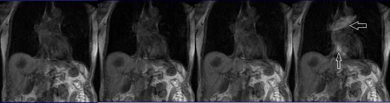
The care bolus technique is the most commonly used method for bolus detection. This technique involves employing a coronal fast gradient refocused sequence to obtain real-time images every second through the vascular structure of interest, typically positioned over the heart. By monitoring the arrival of the contrast bolus in the heart, the operator can then switch to the centric 3D dynamic sequence for further imaging.
Planning care bolus
Plan the coronal care bolus slice on the sagittal plane. Position the block over the mid-heart and angle the slice parallel to the ascending aorta. Verify the position in the other two planes. Establish an appropriate angle in the axial plane, aligning it vertically across the liver.
Care bolus scans should commence one second before contrast administration. The operator can then observe the scans in real-time and monitor the arrival of the contrast bolus in the heart. Once the contrast reaches the heart, the care bolus should be promptly halted, and the patient should be instructed to hold their breath before initiating the centric 3D dynamic sequence.
T1 vibe dixon\ flash 3D fat sat axial breath hold dynamic 2 RUN
Plan the axial slices on the coronal plane; angle the position block parallel to the right and left renal pelvis. Check the positioning block in the other two planes. An appropriate angle must be given in the sagittal plane (perpendicular to the long axis of kidney). Slices must be sufficient to cover both kidneys from two slices above the upper pole of kidneys down to two slices below the lower pole of kidney. Phase oversampling and, in the case of 3D blocks, slice oversample, must be used to avoid wrap around artefacts. Consider adding saturation bands at the top and bottom of the block to minimize artifacts caused by fat signal, arterial pulsation, and breathing. Instruct the patient to hold their breath during image acquisition.
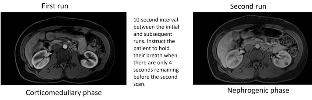
A dynamic VIBE Dixon 3D sequence comprises two VIBE 3mm 3D scans with a 10-second delay between the first and second scans. The first scan captures the arterial phase, while the second scan captures the venous phase. Timing for each scan is crucial. Proper breathing instructions should be given during both scans. After the arterial phase scan, the patient should be instructed to breathe normally, and then asked to hold their breath when there are approximately 4 seconds remaining for the venous scan.
Alternatively, the post-contrast dynamic scans can be performed by splitting the sequence into two separate scans and conducting them individually according to their respective timelines. In our department, we prefer to perform them separately.
Some radiologists prefer to generate subtraction images between the pre-contrast and post-contrast images. To enable image subtraction, it is essential that the measurements of pre- and post-contrast T1 scans are identical. Any variations in slice thickness, field of view (FOV), or planning would impede the ability to perform image subtraction.
Parameters FLASH
| TR 4-5 | TE 2 | FAT SAT ON | NEX 1 | SLICE 3MM | MATRIX 320X320 | FOV 350 | PHASE A>P | DYNAMIC 3 SCANS | IPAT ON |
Parameters VIBE DIXON
TR 6-7 | TE 2.39 4.77 | FLIP 10 | NXA 1 | SLICE 3 MM | MATRIX 320×320 | FOV 320-350 | PHASE A>P | OVERSAMPLE 20% | BH YES |
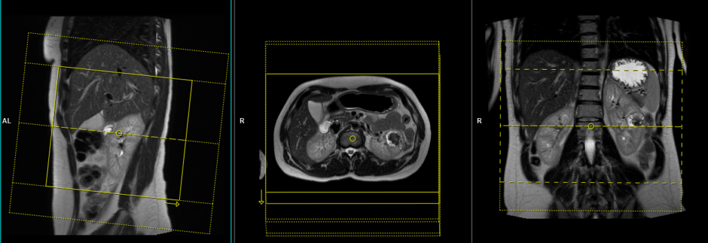
T1 vibe DIXON breath hold coronal 4mm post contrast
Plan the coronal slices on the axial plane and angle the positioning block parallel to the right and left kidneys. Check the positioning block in the other two planes. An appropriate angle must be given in the sagittal plane, parallel to the long axis of the kidneys. Ensure that the slices are sufficient to cover both kidneys from anterior to posterior. Phase oversampling and slice oversampling must be used to avoid wrap-around artifacts. Instruct the patient to hold their breath during image acquisition. In our department, we instruct patients to breathe in and out twice before giving the ‘breathe in and hold’ instruction.
Parameters VIBE dixon
TR 6-7 | TE 2.39 4.77 | FLIP 10 | NXA 1 | SLICE 4 MM | MATRIX 320×320 | FOV 320-350 | PHASE A>P | OVERSAMPLE 20% | BH YES |