Sacroiliac Joints(SIJ) MRI Protocols and Planning
Indications of sacroiliac joints MRI
- Neoplasms of bone, joint, or soft tissue
- Infections of bone, joint, or soft tissue
- Inflammation of the Sacroiliac joint
- Sacroiliac joint dysfunction
- ankylosing spondylitis
- Degenerative arthritis
- Sacroiliac joint pain
- Avascular necrosis
- Rheumatoid arthritis
- Osteoarthritis
- Sacroiliitis
- Psoriasis
- Gout
Contraindications
- Any electrically, magnetically or mechanically activated implant (e.g. cardiac pacemaker, insulin pump biostimulator, neurostimulator, cochlear implant, and hearing aids)
- Intracranial aneurysm clips (unless made of titanium)
- Pregnancy (risk vs benefit ratio to be assessed)
- Ferromagnetic surgical clips or staples
- Metallic foreign body in the eye
- Metal shrapnel or bullet
Patient preparation for sacroiliac joints MRI
- A satisfactory written consent form must be taken from the patient before entering the scanner room
- Ask the patient to remove all metal object including keys, coins, wallet, any cards with magnetic strips, jewellery, hearing aid and hairpins
- Ask the patient to undress and change into a hospital gown
- If possible, provide a chaperone for claustrophobic patients (e.g. relative or staff )
- Offer earplugs or headphones, possibly with music for extra comfort
- Explain the procedure to the patient
- Instruct the patient to keep still
- Note the weight of the patient
Positioning for sacroiliac joints MRI
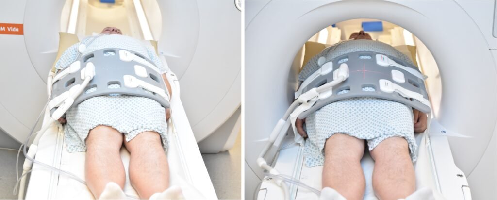
- Position the patient in supine position with head pointing towards the magnet (head first supine)
- Position the patient over the spine coil and place the body coil over the pelvis(three inches above iliac crest down to symphysis pubis)
- Securely tighten the body coil using straps to prevent respiratory artefacts
- Give a pillow under the head for extra comfort (do not give cushions under the legs )
- Centre the laser beam localiser over anterior superior iliac spine (2 inches below iliac crest)
Suggested protocols, parameters and planning
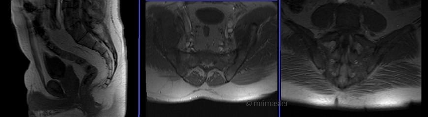
sacroiliac joints MRI Localizer
A three-plane localizer must be taken at the beginning to localize and plan the sequences. Localizers typically take less than 25 seconds and are T1-weighted with low resolution.
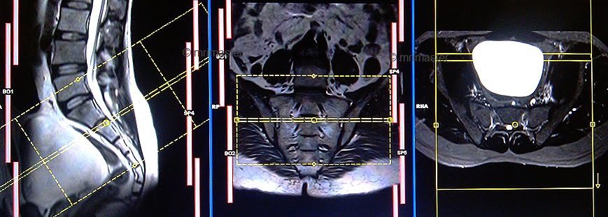
T2 stir coronal oblique 3mm SFOV
Plan the coronal oblique slices on the sagittal plane; angle the positioning block parallel to the sacrum. Check the positioning block in the other two planes. An appropriate angle must be given in the axial plane (parallel to the sacral ala). Slices must be sufficient to cover the whole sacroiliac joints. The phase direction must be right to left with 100% oversampling to avoid wrap-around artifacts from breathing.
Parameters
TR 4000-5000 | TE 110 | FLIP 150 | NEX 2 | SLICE 3MM | MATRIX 320X256 | FOV 200-270 | PHASE R>L | GAP 10% | TI 150 |
T1 tse coronal oblique 3mm SFOV
Plan the coronal oblique slices on the sagittal plane; angle the positioning block parallel to the sacrum. Check the positioning block in the other two planes. An appropriate angle must be given in the axial plane (parallel to the sacral ala). Slices must be sufficient to cover the whole sacroiliac joints. The phase direction must be right to left with 100% oversampling to avoid wrap-around artifacts from breathing.
Parameters
TR 400-600 | TE 15-25 | SLICE 3 MM | FLIP 150 | PHASE R>L | MATRIX 320X320 | FOV 200-270 | GAP 10% | NEX(AVRAGE) 2 |
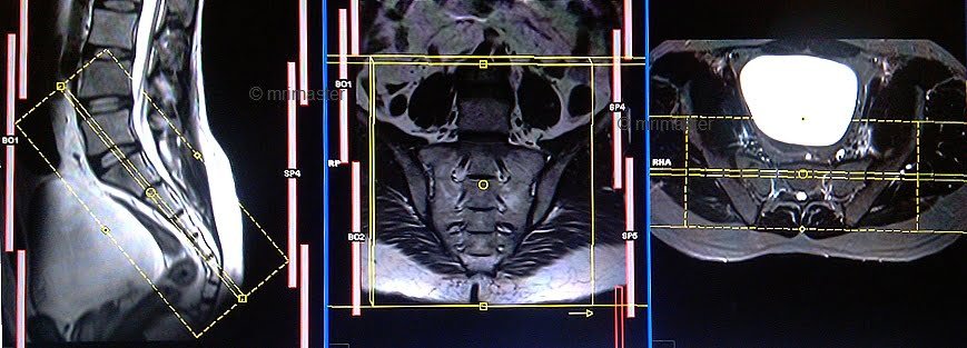
T2 stir axial oblique 3mm SFOV
Plan the axial oblique slices on the sagittal plane; angle the positioning block perpendicular to the sacrum. Check the positioning block in the other two planes. An appropriate angle must be given in the coronal plane (perpendicular to the sacrum). Slices must be sufficient to cover the whole sacroiliac joints. Adding saturation bands in front of the axial block will reduce artifacts from breathing and peristalsis.
Parameters
TR 4000-5000 | TE 110 | FLIP 160 | NEX 2 | SLICE 3MM | MATRIX 320X256 | FOV 200-270 | PHASE R>L | GAP 10% | TI 150 |

